


Mega Doctor News
Newswise — NIBIB-funded engineers at the University of Buffalo have fine-tuned the use of stereolithography for 3D printing of organ models that contain live cells. The new technique is capable of printing the models 10-50 times faster than the industry standard—in minutes instead of hours— a major step in the quest to create 3D-printed replacement organs.
Conventional 3D printing involves the meticulous addition of material to the 3D model with a small needle that produces fine detail but is extremely slow –taking six or seven hours to print a model of a human part, such as a hand, for instance. The lengthy process causes cellular stress and injury inhibiting the ability to seed the tissues with live, functioning cells.
The method developed by the SUNY Buffalo group, led by Rougang Zhao, PhD, Associate Professor of Biomedical Engineering in the Jacobs School of Medicine & Biomedical Sciences, takes a different approach that minimizes damage to live cells. The rapid, cell friendly technique is a significant step towards creating printed tissues infused with large numbers of living cells.


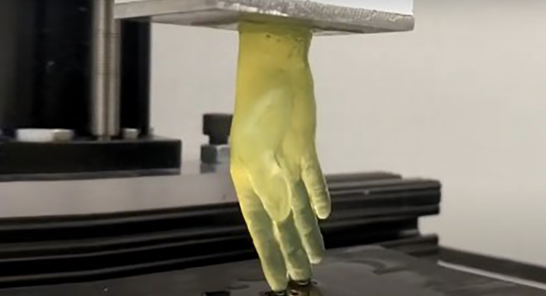
Instead of a needle moving across a surface slowly building the details of the 3D model, Zhao and his team members have developed a system that enables a small 10 centimeter model of a human hand, to rise—quite dramatically—out of a vat of liquid in a matter of minutes, instead of hours (see video). The stereolithographic method employs projected light at the bottom of the tank that penetrates up through the mix of hydrogel and live cells. The light polymerizes the hydrogel/cell mixture at precise positions in the model, building entire layers of the model continually—rather than one painstaking pinpoint at a time.
“This is a significant step towards the printing of biologically active 3D tissues,” explained David Rampulla, Ph.D., director of the NIBIB program in Synthetic Biological Systems. “This new technique combines a hydrogel mix that is very cell friendly with the rapid printing process, which spares cells from being suspended in a cell-damaging environment for an extended period. The combination has allowed the team to introduce live cells into their 3D printed tissues with the vast majority of cells remaining alive and functional.”
With an eye on moving from small models of organs to full-sized replacement organs in the future, the engineering team successfully used the new method to print tissues containing inner branching networks that mimic blood vessels. “To keep alive cells that are deep inside a full-sized printed organ is one of the many challenges in creating functional replacement organs,” explained Zhao. “We found that our method performed well in terms of creating branched, vessel-like networks to facilitate delivery of nutrients to cells embedded throughout the printed tissue.”
Going a step further, the team then introduced live endothelial cells into the branching canals of their 3D-printed tissues. The endothelial cells adhered to the walls of the artificial vasculature and expanded to form an endothelial lining similar to that found in actual blood vessels.
The research team is now working to increase the size of the printed tissues while maintaining structural integrity and cell viability. The work is a significant step towards the lofty, yet potentially obtainable goal of many bioengineers—printing replacement organs—a biomedical advance that could save countless lives lost because of the pervasive shortage of donor organs, such as kidneys and livers.
This study was supported by National Institute of Biomedical Imaging and Bioengineering of the National Institutes of Health under award number R01EB019411. The authors also acknowledge the funding support from the School of Engineering and Jacobs School of Medicine and Biomedical Sciences at the University at Buffalo. The work was reported in Advanced Healthcare Materials.